Describe Image Formation on the Retina
Degenerative diseases of the human retina. Thrombus and embolus are two terms used interchangeably to describe blood clots.
Physics Of The Eye Physics Ii
While the indication for treatment.
. 95 the two right-angled triangles ABF and. In some eye diseases the retina becomes damaged or compromised and degenerative changes set in that eventally lead to serious damage to the nerve cells that carry the vital mesages about the visual image to. A thrombus in a vein is.
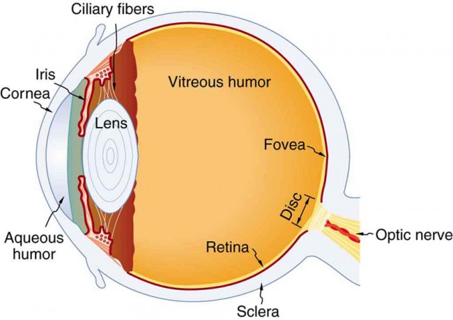
Rete net is the innermost light-sensitive layer of tissue of the eye of most vertebrates and some molluscsThe optics of the eye create a focused two-dimensional image of the visual world on the retina which translates that image into electrical neural impulses to the brain to create visual perceptionThe retina serves a function analogous to that of the film or. A dedicated set of retinal ganglion cells RGCs and brainstem visual nuclei termed the accessory optic system AOS generate slip-compensating eye movements that stabilize visual images on the retina and improve visual performance. When a red cross passed across the screen about one third of the subjects did not notice it.
The retina actually consists of two components. Which types of RGCs project to each of the various AOS nuclei. Refractive indices are crucial to image formation using lenses.
A second target of the axons of neurons in the vestibular nuclei is the spinal cord which initiates the spinal reflexes involved with posture and balance. An outermost layer of retinal pigment epithelium RPE which is composed of single layer of cuboidal melanin-containing cells and the neural retina which is a multilayered structure containing photoreceptors as well as neurons and glia. Somewhat paradoxically the optical properties of the eye that allow image formation prevent direct inspection of the retina.
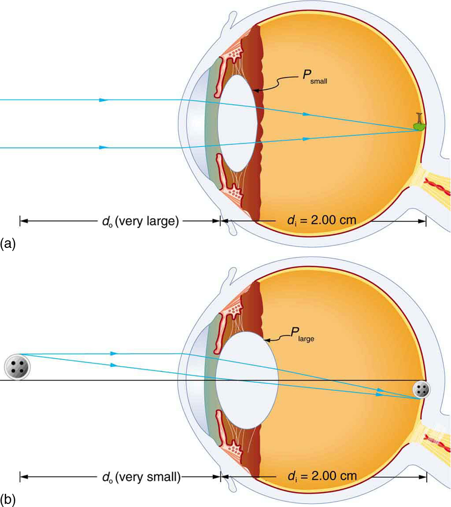
Table 164 shows refractive indices relevant to the eye. When the head rotates the image of the visual world slips across the retina. The biggest change in the refractive indexand the one that causes the greatest bending of raysoccurs at the cornea rather than the lens.
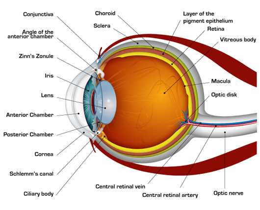
In other words the very nature of the imaging transform resulting in a focused image on the retinal surface disallows depiction of the retina when attempting to form a focused retinal image from the outside via usage of the inverse. In life these two components are fused into what we typically call the retina and it is subdivided. Gap junctions are specialized intercellular connections between a multitude of animal cell-types.
Thus point A is image point of A if every ray originating at point A and falling on the concave mirror after reflection passes through the point A. To assist the visual system fibers of the vestibular nuclei project to the oculomotor trochlear and abducens. We now derive the mirror equation or the relation between the object distance u image distance v and the focal length f.
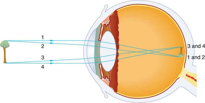
Participants were instructed to focus on either white or black objects disregarding the other color. The ray diagram in Figure 1633 shows image formation by the cornea and lens of. This point is called the focal point.
The retina from Latin. Perhaps the most common example of photothermal damage to the retina is in the form of the clinical usage of lasers for the treatment of various disease states of the retina including diabetic retinopathy retinal oedema retinopathy of prematurity tumours of the choroid and retina retinal tears and retinal detachments Figure 4. The formation of blood clots is induced by certain conditions such as high cholesterol tobacco smoking diabetes cancer being obese or overweight stress and sedentary lifestyle.
The human retina is a delicate organization of neurons glia and nourishing blood vessels. They directly connect the cytoplasm of two cells which allows various molecules ions and electrical impulses to directly pass through a regulated gate between cells. Aggregation of platelets forms a quick plug to prevent bleeding.
Everybody who ever misused a magnifying glass as a burning glass has discovered that a lens creates a hot spot when pointed at the sun. In other words an image focuses on the retina nerve cells process the information and they send it by electrical impulses to the brain. One gap junction channel is composed of two protein hexamers or hemichannels called connexons in.
Image formation through a lens Before exploring how such a lens works crucial terms and definitions of a lens have to be clarified. One target is the reticular formation which influences respiratory and cardiovascular functions in relation to body movements. Weblio辞書 - retina とは意味目の網膜.
例文a part of an eye called retina. What concept is illustrated by the following study. The distance from the center of the lens to this focal point.
Physics Of The Eye Physics Ii
How The Human Eye Works Cornea Layers Role Light Rays
What Is An Image Behind The Retina Quora
Physics Of The Eye Physics Ii


Comments
Post a Comment